0091 98 967 44 968
dineshvrm@gmail.com
Newsletter
0091 98 967 44 968
dineshvrm@gmail.com
Menu
Categories
- Refurbished
- Home Decor & Art
- Showpieces
- Homekeeping
- Chemistry Lab
- Showpieces
- Pharmacology Lab
- Housekeeping
- Ovens & Incubators
- ICU Beds
- Tableware
- Metallurgical
- Tool Maker's Microscope
- Civil Lab
- Specimen Mounting Press
- Brinell Microscope
- Jominy End Quench
- Specimen Leveller
- Metallurgical Microscopes
- Muffle Furnace
- Spectro Polisher
- Specimen Cutting Machine
- Autocollimator
- Specimen Sets
- Polishing Machine
- Material Pro Software
- Belt Grinder
- Vickers Hardness Tester
- Profile Projector
- Rockwell Hardness Tester
- Microscopes
- Penta Head Microscopes
- Laboratory Microscopes
- Stereo Microscope
- Polarising & ORE Microscopes
- Deca Head Microscope
- INDUSTRIAL MICROSCOPES
- Travelling Microscope
- Dissecting and Student Microscope
- Ultra Series Microscope
- Medical and Research
- Inverted Tissue Culture
- Fluorescence Mcroscope
- Projection Microscope
- Digital Microscope
- Houseware & Room
- Laboratory Glassware
- Fowler Bed
- Vintage Decorative
- Indoor & Outdoor
- SEMI FOWER BED
- Wall Accent
- Indoor Plants
- Plain Bed
- Wall Accents
- Biology Lab
- Lighting & Lamps
- Delivery Beds & Tables
- Physics Lab
- Lighting & Lamps
- Laboratory
- Mattress & Accent
- Softwares
- Mattress Topper
- MICROPHOTOGRAPHY
Wishlist
Please, install YITH Wishlist plugin
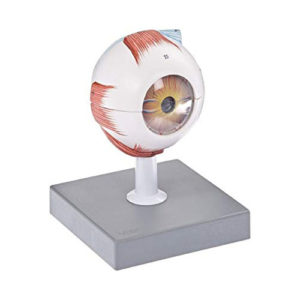
Human Eye 6 Parts Anatomy Model
Dissectible into 6 parts, showing both halves of sclera with cornea and eye muscle attachments and choroid with iris and retina, lens, vitreous humor mounted on a stand with numbered key card.
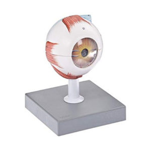
Human Eye 6 Parts Anatomy Model
Dissectible into 6 parts, showing both halves of sclera with cornea and eye muscle attachments and choroid with iris and retina, lens, vitreous humor mounted on a stand with numbered key card.
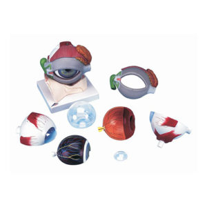
Human Eye Demonstration Model
One side of this 6x model shows the eye socket with a sagittal cut away, while the other side of the model shows the outside of the eye and all the muscle attachments, as well as illustrations depicting how the eye focuses. The cut away is encased in a clear protective covering and two separate insets show the fine structure of the retina as seen with an electron microscope, on a base with Key Card.
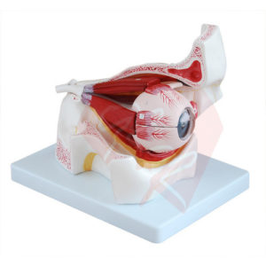
Human Eye Enlarged 5 Times Anatomy Model
Eye model 5x enlarged, dissectible into 6 parts, showing both halves of sclera with cornea and eye muscle attachments and choroid with iris and retina, lens, vitreous humor mounted on a stand with numbered key card.
Human Eye Enlarged 5 Times Anatomy Model
Eye model 5x enlarged, dissectible into 6 parts, showing both halves of sclera with cornea and eye muscle attachments and choroid with iris and retina, lens, vitreous humor mounted on a stand with numbered key card.
Human Eye with Eyelids Anatomy Model
Dissectible into 8 parts, 5 Times enlarged model includes eyelids showing the bony orbit with intraocular muscles, a tear duct, and lachrymal gland. Cornea, muscles, 2 part choroid with iris, retina, and optic nerve; clear lens; and clear vitreous humor are removable, mounted on a stand with key card.
Human Eye with Orbit Anatomy Model
Dissectible into 7 parts, 4X life size, shows the eyeball with optic nerves and muscles in its natural position in the bony orbit, mounted on a stand with numbered key card.
Human Eye with Orbit Anatomy Model
Dissectible into 7 parts, 4X life size, shows the eyeball with optic nerves and muscles in its natural position in the bony orbit, mounted on a stand with numbered key card.
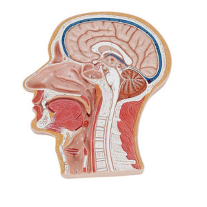
Human Head and Neck Anatomy Model
Life size representation of the superficial and the internal structures of the head and neck. This relief model shows all relevant structures of the human head in great detail. Mounted on board. With Key Card.
Human Head and Neck Anatomy Model
Life size representation of the superficial and the internal structures of the head and neck. This relief model shows all relevant structures of the human head in great detail. Mounted on board. With Key Card.
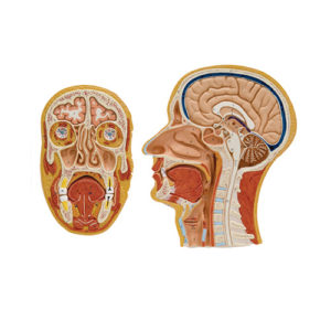
Human Head, Median and Frontal Section Anatomy Model
Life size representation of the superficial and the internal structures of the head and neck. Comparison between frontal and median sections provides a superior understanding of the anatomical structures of the head and neck, mounted on board with key card.
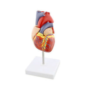
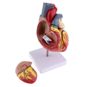
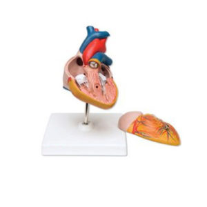
Human Heart 2 Parts Anatomy Model
Life size in 2 parts, anterior heart wall can be removed to show the left and right ventricles and atria as well as the tricuspid, pulmonary, mitral and aortic valves, mounted on base with stand and key card.
Human Heart 2 Parts Anatomy Model
Life size in 2 parts, anterior heart wall can be removed to show the left and right ventricles and atria as well as the tricuspid, pulmonary, mitral and aortic valves, mounted on base with stand and key card.
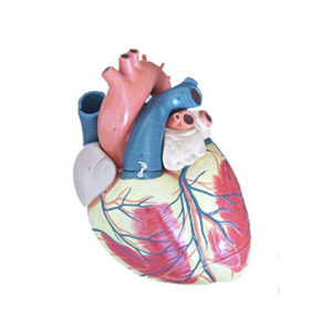
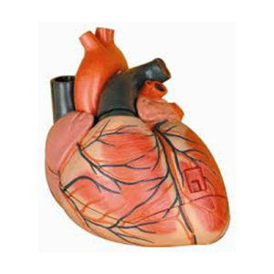
Human Heart 3 Times Life Size Anatomy Model
3 times life size and separates into four parts for detailed study. Sectioned so that both ventricles and atria open to expose the valves & large blood vessels near the heart and musculature of the heart are also shown, mounted on base with Key Card.
Human Heart 3 Times Life Size Anatomy Model
3 times life size and separates into four parts for detailed study. Sectioned so that both ventricles and atria open to expose the valves & large blood vessels near the heart and musculature of the heart are also shown, mounted on base with Key Card.
Human Heart 7 Parts Anatomy Model
This model shows the anatomy of the human heart can be dissectible in 7 parts. The model is mounted on the base and includes a key Card for identifying parts: Oesophagus, Trachea, Superior vena cava, Aorta, Front Heart Wall, Upper half of the heart.
Human Heart 7 Parts Anatomy Model
This model shows the anatomy of the human heart can be dissectible in 7 parts. The model is mounted on the base and includes a key Card for identifying parts: Oesophagus, Trachea, Superior vena cava, Aorta, Front Heart Wall, Upper half of the heart.
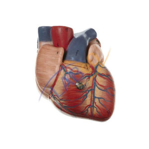
Human Heart Anatomy Model
Dissectible into 2 parts, removable from base, shows internal and external anatomy including valves, cardiac chambers, and pulmonary and systemic vascular structures. Mounted on base, with key card
Human Heart Anatomy Model
Dissectible into 2 parts, removable from base, shows internal and external anatomy including valves, cardiac chambers, and pulmonary and systemic vascular structures. Mounted on base, with key card
Human Heart, 3 Parts Anatomy Model
Human Heart model dissectible in 3 parts, showing, Bicuspid and tricuspid semilunar and sigmoid values on base, with key card.
Human Heart, 3 Parts Anatomy Model
Human Heart model dissectible in 3 parts, showing, Bicuspid and tricuspid semilunar and sigmoid values on base, with key card.
Human Heart, 4 Parts Anatomy Model
Dissectible in 4 parts, this model is enlarged 2 times the life size for a closer look at the intricate inner structure. The model is mounted on base with key card.
Human Heart, 4 Parts Anatomy Model
Dissectible in 4 parts, this model is enlarged 2 times the life size for a closer look at the intricate inner structure. The model is mounted on base with key card.
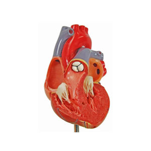
Human Heart, Economy Anatomy Model
Human Heart Economy model dissectible in 2 parts, natural size, showing frontal section and the front of the heart on base, with key card.
Human Heart, Economy Anatomy Model
Human Heart Economy model dissectible in 2 parts, natural size, showing frontal section and the front of the heart on base, with key card.
Human Larynx Anatomy Model
Dissectible in 2 parts, full size, showing muscles, thyroid glands and larynx, mounted on base with key card.
Human Larynx Anatomy Model
Dissectible in 2 parts, full size, showing muscles, thyroid glands and larynx, mounted on base with key card.
Categories
- Biology Lab
- Chemistry Lab
- Delivery Beds & Tables
- Fowler Bed
- Home Decor & Art
- Homekeeping
- Housekeeping
- Houseware & Room
- ICU Beds
- Indoor & Outdoor
- Indoor Plants
- Laboratory
- Laboratory Glassware
- Lighting & Lamps
- Lighting & Lamps
- Mattress & Accent
- Mattress Topper
- Metallurgical
- Autocollimator
- Belt Grinder
- Brinell Microscope
- Civil Lab
- Jominy End Quench
- Material Pro Software
- Metallurgical Microscopes
- Muffle Furnace
- Polishing Machine
- Profile Projector
- Rockwell Hardness Tester
- Specimen Cutting Machine
- Specimen Leveller
- Specimen Mounting Press
- Specimen Sets
- Spectro Polisher
- Tool Maker's Microscope
- Vickers Hardness Tester
- MICROPHOTOGRAPHY
- Microscopes
- Deca Head Microscope
- Digital Microscope
- Dissecting and Student Microscope
- Fluorescence Mcroscope
- INDUSTRIAL MICROSCOPES
- Inverted Tissue Culture
- Laboratory Microscopes
- Medical and Research
- Penta Head Microscopes
- Polarising & ORE Microscopes
- Projection Microscope
- Stereo Microscope
- Travelling Microscope
- Ultra Series Microscope
- Ovens & Incubators
- Pharmacology Lab
- Physics Lab
- Plain Bed
- Refurbished
- SEMI FOWER BED
- Showpieces
- Showpieces
- Softwares
- Tableware
- Uncategorized
- Vintage Decorative
- Wall Accent
- Wall Accents
Product Status
Example Title
Door sit amet, consectetur adip iscing elit, sed do ore magna lorem ipsum sit.
MORE
You may like it
More
More

