- Refurbished
- Home Decor & Art
- Showpieces
- Homekeeping
- Chemistry Lab
- Showpieces
- Pharmacology Lab
- Housekeeping
- Ovens & Incubators
- ICU Beds
- Tableware
- Metallurgical
- Tool Maker's Microscope
- Civil Lab
- Specimen Mounting Press
- Brinell Microscope
- Jominy End Quench
- Specimen Leveller
- Metallurgical Microscopes
- Muffle Furnace
- Spectro Polisher
- Specimen Cutting Machine
- Autocollimator
- Specimen Sets
- Polishing Machine
- Material Pro Software
- Belt Grinder
- Vickers Hardness Tester
- Profile Projector
- Rockwell Hardness Tester
- Microscopes
- Penta Head Microscopes
- Laboratory Microscopes
- Stereo Microscope
- Polarising & ORE Microscopes
- Deca Head Microscope
- INDUSTRIAL MICROSCOPES
- Travelling Microscope
- Dissecting and Student Microscope
- Ultra Series Microscope
- Medical and Research
- Inverted Tissue Culture
- Fluorescence Mcroscope
- Projection Microscope
- Digital Microscope
- Houseware & Room
- Laboratory Glassware
- Fowler Bed
- Vintage Decorative
- Indoor & Outdoor
- SEMI FOWER BED
- Wall Accent
- Indoor Plants
- Plain Bed
- Wall Accents
- Biology Lab
- Lighting & Lamps
- Delivery Beds & Tables
- Physics Lab
- Lighting & Lamps
- Laboratory
- Mattress & Accent
- Softwares
- Mattress Topper
- MICROPHOTOGRAPHY
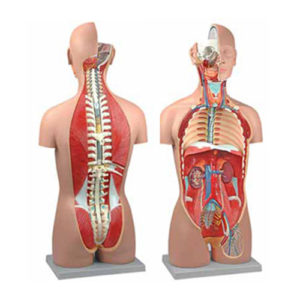
Human Torso, Dual Sex with Open Back 27-Part
This sexless, life-size torso on base is accurate in all of its detailing. Anatomical structures of the major body systems are numbered an identified on the accompany key. The head is sectioned to expose one half of the brain. The neck is dissected through the ventral surface to show muscular, neural, vascular, and glandular structures. The thorax and abdomen are completely open, for an unrestricted view of the internal organs and associated structures. The back is dissected, revealing the muscular layers, vertebral column, spinal cord, and nerve branches. One thoracic vertebra is removable for detailed examination of this structure and associations with the spinal cord.
Lobule of the Lung Anatomy Model
Lobule of the Lung Anatomy Model
Neuron Anatomy Model
Neuron Anatomy Model
Categories
- Biology Lab
- Chemistry Lab
- Delivery Beds & Tables
- Fowler Bed
- Home Decor & Art
- Homekeeping
- Housekeeping
- Houseware & Room
- ICU Beds
- Indoor & Outdoor
- Indoor Plants
- Laboratory
- Laboratory Glassware
- Lighting & Lamps
- Lighting & Lamps
- Mattress & Accent
- Mattress Topper
- Metallurgical
- Autocollimator
- Belt Grinder
- Brinell Microscope
- Civil Lab
- Jominy End Quench
- Material Pro Software
- Metallurgical Microscopes
- Muffle Furnace
- Polishing Machine
- Profile Projector
- Rockwell Hardness Tester
- Specimen Cutting Machine
- Specimen Leveller
- Specimen Mounting Press
- Specimen Sets
- Spectro Polisher
- Tool Maker's Microscope
- Vickers Hardness Tester
- MICROPHOTOGRAPHY
- Microscopes
- Deca Head Microscope
- Digital Microscope
- Dissecting and Student Microscope
- Fluorescence Mcroscope
- INDUSTRIAL MICROSCOPES
- Inverted Tissue Culture
- Laboratory Microscopes
- Medical and Research
- Penta Head Microscopes
- Polarising & ORE Microscopes
- Projection Microscope
- Stereo Microscope
- Travelling Microscope
- Ultra Series Microscope
- Ovens & Incubators
- Pharmacology Lab
- Physics Lab
- Plain Bed
- Refurbished
- SEMI FOWER BED
- Showpieces
- Showpieces
- Softwares
- Tableware
- Uncategorized
- Vintage Decorative
- Wall Accent
- Wall Accents
Product Status

Example Title
Door sit amet, consectetur adip iscing elit, sed do ore magna lorem ipsum sit.


