0091 98 967 44 968
dineshvrm@gmail.com
Newsletter
0091 98 967 44 968
dineshvrm@gmail.com
Menu
Categories
- Refurbished
- Home Decor & Art
- Showpieces
- Homekeeping
- Chemistry Lab
- Showpieces
- Pharmacology Lab
- Housekeeping
- Ovens & Incubators
- ICU Beds
- Tableware
- Metallurgical
- Tool Maker's Microscope
- Civil Lab
- Specimen Mounting Press
- Brinell Microscope
- Jominy End Quench
- Specimen Leveller
- Metallurgical Microscopes
- Muffle Furnace
- Spectro Polisher
- Specimen Cutting Machine
- Autocollimator
- Specimen Sets
- Polishing Machine
- Material Pro Software
- Belt Grinder
- Vickers Hardness Tester
- Profile Projector
- Rockwell Hardness Tester
- Microscopes
- Penta Head Microscopes
- Laboratory Microscopes
- Stereo Microscope
- Polarising & ORE Microscopes
- Deca Head Microscope
- INDUSTRIAL MICROSCOPES
- Travelling Microscope
- Dissecting and Student Microscope
- Ultra Series Microscope
- Medical and Research
- Inverted Tissue Culture
- Fluorescence Mcroscope
- Projection Microscope
- Digital Microscope
- Houseware & Room
- Laboratory Glassware
- Fowler Bed
- Vintage Decorative
- Indoor & Outdoor
- SEMI FOWER BED
- Wall Accent
- Indoor Plants
- Plain Bed
- Wall Accents
- Biology Lab
- Lighting & Lamps
- Delivery Beds & Tables
- Physics Lab
- Lighting & Lamps
- Laboratory
- Mattress & Accent
- Softwares
- Mattress Topper
- MICROPHOTOGRAPHY
Wishlist
Please, install YITH Wishlist plugin
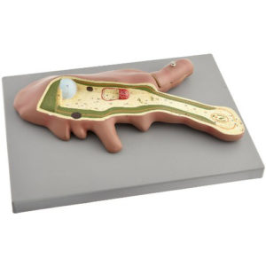
ABSORPTION ZONE OF ROOT
Model of white mustard (sinapis alba) showing the absorption zone of a dicotledonous plant, mounted on base with key card.
ABSORPTION ZONE OF ROOT
Model of white mustard (sinapis alba) showing the absorption zone of a dicotledonous plant, mounted on base with key card.
AIDS VIRUS MODEL
Model of HIV virus is enlarged millions of times shows the outer lipid membrane with protein structures, and the internal nucleus which contains the viral hereditary matter (RNA). The nucleus is removable and condoms can be put underneath to provide a message regarding measures to take in protecting against HIV infections, mounted on base with key card.
Amoeba Model – Zoology Anatomy Model
Zoology Model Amoeba proteus, enlarged approx. 1000 times. In a small pseudopodium which can be opened up showing the structure after electron microscopic magnification, mounted on base with key card.
Amoeba Model – Zoology Anatomy Model
Zoology Model Amoeba proteus, enlarged approx. 1000 times. In a small pseudopodium which can be opened up showing the structure after electron microscopic magnification, mounted on base with key card.
Anatomy of the Foot Anatomy Model
Life size model, dissectible in 3 parts. The plantar aponeurosis and the flexor brevis can be removed to show the underlying network of muscles, tendons, vessels and nerves. A deeper plantar dissection allows observation of the plantar muscles and plantar nerve branches. A superficial dorsal dissection shows ligaments, nerves and vessels.
Animal Cell Division Meiosis Zoology Model
Animal Cell Division Meiosis Zoology Model - Set of 12 models, showing detailed stages in Meiosis cell division, beautifully coloured to bring out the detailed structure as seen under the microscope, mounted on base with key card.
Animal Cell Division Meiosis Zoology Model
Animal Cell Division Meiosis Zoology Model - Set of 12 models, showing detailed stages in Meiosis cell division, beautifully coloured to bring out the detailed structure as seen under the microscope, mounted on base with key card.
Animal Cell Division Mitosis Zoology Model
Animal Cell Division Mitosis - A set of 10 models, showing resting cell, early prophase, prophase, late prophase, metaphase, anaphase, late anaphase, Telophase and daughter cells, mounted on base with key card.
Animal Cell Division Mitosis Zoology Model
Animal Cell Division Mitosis - A set of 10 models, showing resting cell, early prophase, prophase, late prophase, metaphase, anaphase, late anaphase, Telophase and daughter cells, mounted on base with key card.
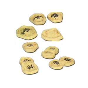
Animal Cell Mitosis Zoology Model Set of 8
Robust Models illustrating the process of mitotic cell division. The chromosomes are painted to allow easy identification, mounted on base with key card.
Animal Cell Mitosis Zoology Model Set of 8
Robust Models illustrating the process of mitotic cell division. The chromosomes are painted to allow easy identification, mounted on base with key card.
Animal Cell Three Dimensional Zoology Anatomy Model
Animal Cell Three Dimensional Zoology Anatomy Model - A three dimensional model with one side dissected to show complete internal structure, mounted on base with key card.
Animal Cell Three Dimensional Zoology Anatomy Model
Animal Cell Three Dimensional Zoology Anatomy Model - A three dimensional model with one side dissected to show complete internal structure, mounted on base with key card.
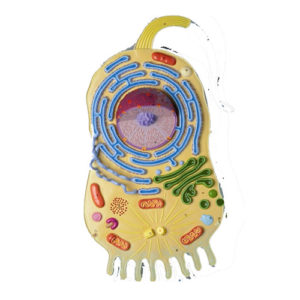
Animal Cell Ultra-Structure Zoology Model
Model of cell showing ultrastructure, mounted on base with key card.
Animal Cell Ultra-Structure Zoology Model
Model of cell showing ultrastructure, mounted on base with key card.
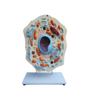
Animal Cell Zoology Model
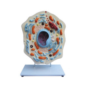
This model, enlarged about 20,000 times. Animal Cell Model, provides clear depiction of the delicate and complex structure of the animal cell. Shows the nucleus , endoplasmatic reticulum, mitochondria, ribosomes, polysomes and Golgi apparatus, centrioles, lysosomes and fat vacuoles.
ASCARIS MALE AND FEMALE
Enlarged, dissection showing the internal organs of both male and female, mounted on base with key card.
ASCARIS MALE AND FEMALE
Enlarged, dissection showing the internal organs of both male and female, mounted on base with key card.
Cat Dissection Model
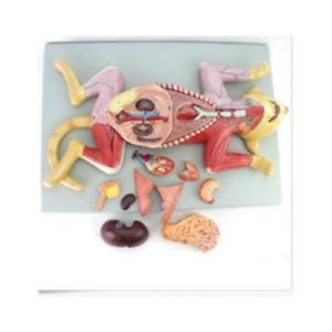
This life-size dissection model features over 100 individual anatomical details. An extremely realistic and precise model, it is crafted with hand-painted detail. Featured structures include a cross-sectioned kidney showing the cortex and medulla, major arteries and veins, muscle groups of the fore and hind limbs, and the open uterus exposing a developing fetus. This model also has an open mouth cavity detailing the teeth and nasopharynx, and includes a key identifying 136 structures.
COMPARATIVE GERMINATION MODEL
Eight-piece model for comparison of three different types of germination methods: monocot, dicot and gymnosperm.
Using suitable models from the plant world, students can study root development, sprouting methods and the structure of the seedling. Each component of the model is removable to facilitate closer examination, mounted on base with key card.
Dental Hygiene Anatomy Model
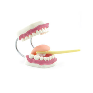
3x life size model, useful for teaching to brush teeth including giant toothbrush on base with key card.
Dental Hygiene Anatomy Model
3x life size model, useful for teaching to brush teeth including giant toothbrush on base with key card.
DICOT LEAF V.S.
Showing details of typical mesophytic leaf, mounted on base with key card.
DICOT LEAF V.S.
Showing details of typical mesophytic leaf, mounted on base with key card.
Categories
- Biology Lab
- Chemistry Lab
- Delivery Beds & Tables
- Fowler Bed
- Home Decor & Art
- Homekeeping
- Housekeeping
- Houseware & Room
- ICU Beds
- Indoor & Outdoor
- Indoor Plants
- Laboratory
- Laboratory Glassware
- Lighting & Lamps
- Lighting & Lamps
- Mattress & Accent
- Mattress Topper
- Metallurgical
- Autocollimator
- Belt Grinder
- Brinell Microscope
- Civil Lab
- Jominy End Quench
- Material Pro Software
- Metallurgical Microscopes
- Muffle Furnace
- Polishing Machine
- Profile Projector
- Rockwell Hardness Tester
- Specimen Cutting Machine
- Specimen Leveller
- Specimen Mounting Press
- Specimen Sets
- Spectro Polisher
- Tool Maker's Microscope
- Vickers Hardness Tester
- MICROPHOTOGRAPHY
- Microscopes
- Deca Head Microscope
- Digital Microscope
- Dissecting and Student Microscope
- Fluorescence Mcroscope
- INDUSTRIAL MICROSCOPES
- Inverted Tissue Culture
- Laboratory Microscopes
- Medical and Research
- Penta Head Microscopes
- Polarising & ORE Microscopes
- Projection Microscope
- Stereo Microscope
- Travelling Microscope
- Ultra Series Microscope
- Ovens & Incubators
- Pharmacology Lab
- Physics Lab
- Plain Bed
- Refurbished
- SEMI FOWER BED
- Showpieces
- Showpieces
- Softwares
- Tableware
- Uncategorized
- Vintage Decorative
- Wall Accent
- Wall Accents
Product Status

Example Title
Door sit amet, consectetur adip iscing elit, sed do ore magna lorem ipsum sit.
MORE
You may like it
More
More


