


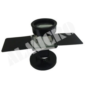
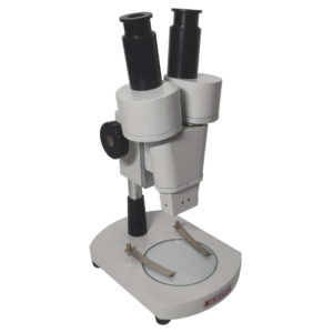
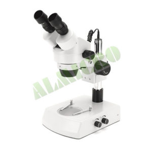
MICROSCOPES-PATHOLOGICAL AND RESEARCH
MAKE DINESH SCIENTIFIC
DESCRIPTION
Because they allow researchers and scientists to examine and examine tissues, cells, and other biological material at a microscopic level, microscopes are essential tools in pathology and medical science. Research and pathology microscopes are made to specifically address the needs of the medical, biological, and allied sciences sectors. The following are some essential elements of using microscopes in research and pathology settings:
for brightfield and phase contrast imaging, which allows for high-resolution viewing of materials, cells, and tissues. The device should have the right light source, high resolution corrected objectives, and the required working distance.
LIGHT MICROSCOPES:
The most popular kind of microscope used in pathology and research are brightfield microscopes. They are useful for seeing stained specimens and offer a bright background.
PHASE-CONTRAST MICROSCOPES:
When viewing translucent or unstained materials, such living cells, phase-contrast microscopy is especially helpful. It improves these specimens’ contrast without the requirement for staining.
FLUORESCENCE MICROSCOPES:
Research uses fluorescence microscopy extensively.
It entails labeling certain molecules or structures within cells with fluorescent dyes or proteins to enable fine-grained imaging of individual cell components.
PATHOLOGY DIGITAL:
Digital imaging technology is used in the collection, organization, and analysis of pathology data in digital pathology. Pathologists can see and evaluate digital slides using whole-slide imaging, which facilitates remote diagnosis and teamwork.
MECHANIZED MICROSCOPY:
Microscopy technologies are becoming more and more automated, enabling higher throughput analysis and more productivity in pathology and research labs.
RESEARCH USES:
In several scientific fields, such as cell biology, microbiology, neuroscience, and developmental biology, microscopes are essential instruments. The structure and operation of cells, tissues, and organisms are studied by researchers using microscopes.
SOFTWARE:
ppropriate software for full image acquisition is supported by the system. Software for image processing and analysis workflows must be simple to use.
Interactive measures include scale bars, length, angles, and contours. The software should support at least the following image formats: BMP, GIF, JPG, and TIFF, as well as the administration, visualization, and printing of metadata and images.
Image stitching, Extended Depth of Focus (EDF) or a similar feature for improved image quality, measurement and annotation tools, and automated fluorescent image acquisition are examples of analysis features that the system should offer in imaging software.
TECHNICAL DETAILS:
MODEL DS-TM-100
MICROSCOPE DESIGN AND BODY
Ergonomic Design stand with manual encoded or motorized focusing
Microscope Body Integrated power unit
Microscope Stand 15 mm Z focus range
Energy Saving (ECO Mode) Automatic switch-off of illumination after 15 minutes of inactivity
Image Capture Snap button available on the microscope stand for direct image capture
LIGHT SOURCE
Light Source 10W LED illumination
Lifespan 60,000 hours
Automatic Light Intensity Yes
Brightness Uniformity Uniform brightness across all magnifications
TRINOCULAR TUBE
Trinocular Tube Binocular phototube with a 30° viewing angle and field diameter of 22 mm
Light Path Distribution Typically includes 100:0 / 0:100 mode
Mechanical Stage 75×50 mm
Objectives
• Both transmitted light and phase contrast applications supported by the system’s objectives.
• The very of the following goals is necessary:
• Achromat 5x/0.12 Plan
• Phase Plan Achromat 10x/0.25
• Plan Achromat 20x/0.45 Phase Plan Achromat 40x/0.65 Phase Plan Achromat 100x/1.25 Phase Abbe condenser of 0.9 for Brightfield application of phase contrast.
Nosepiece The microscope include encoded or motorized revolving nosepiece with 5 objective positions for seamless image capturing with out any loss of the information.
EYEPIECE
Eyepiece 10x eyepiece with a field of view (FOV) of 22 mm
Diopter Available on both eyepieces
Interpupillary Distance 53-74 mm
IMAGING ATTACHMENT
Sensor Type 12MP CMOS with rolling shutter technology
Imaging Mode Colour camera for brightfield and phase contrast imaging
Resolution 4032 (H) x 3044 (V) pixels
Pixel Size 1.85 µm x 1.85 µm or larger at resolution 4032 (H) x 3044 (V) µm
Chip Size Diagonal 9.3 mm (1 / 1.7”) at full sensor
Frame Rate 30 frames per second at resolution
Digitization 3 x 8-bit per pixel
Connectivity Wi-Fi setup or router-based connection
Software Control Operable via Wi-Fi devices (iPad, Laptops, Android mobile phone)
Mount Adapter Suitable C-mount adapter to fit the camera
Optical Reduction zoom adapter desired


Reviews
There are no reviews yet.