
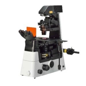


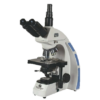
MICROSCOPES PATHOLOGICAL AND RESEARCH
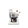
MAKE DINESH SCIENTIFIC MODEL: DS-TM-100
DESCRIPTION:
Because they allow researchers and scientists to examine and examine tissues, cells, and other biological material at a microscopic level, microscopes are essential tools in pathology and medical science. Research and pathology microscopes are made to specifically address the needs of the medical, biological, and allied sciences sectors. The following are some essential elements of using microscopes in research and pathology settings:
LIGHT MICROSCOPES:
The most popular kind of microscope used in pathology and research are brightfield microscopes. They are useful for seeing stained specimens and offer a bright background.
PHASE-CONTRAST MICROSCOPES:
When viewing translucent or unstained materials, such living cells, phase-contrast microscopy is especially helpful. It improves these specimens’ contrast without the requirement for staining.
FLUORESCENCE MICROSCOPES:
Research uses fluorescence microscopy extensively. It entails labeling certain molecules or structures within cells with fluorescent dyes or proteins to enable fine-grained imaging of individual cell components.
PATHOLOGY DIGITAL:
Digital imaging technology is used in the collection, organization, and analysis of pathology data in digital pathology. Pathologists can see and evaluate digital slides using whole-slide imaging, which facilitates remote diagnosis and teamwork.
MECHANIZED MICROSCOPY:
Microscopy technologies are becoming more and more automated, enabling higher throughput analysis and more productivity in pathology and research labs.
RESEARCH USES:
In several scientific fields, such as cell biology, microbiology, neuroscience, and developmental biology, microscopes are essential instruments. The structure and operation of cells, tissues, and organisms are studied by researchers using microscopes.
TECHNICAL DETAILS:
Instrument Type Upright fluorescence microscope
Intended Use Intended for imaging specimens with fluorescent labels.
Applications Ideal for protein localization research in both fixed and living samples, immunofluorescence, live-cell imaging, and FISH
Optical Features Outfitted with the required working distance and high-resolution corrected targets
Illumination & Filters Includes appropriate light source and filters for fluorescence applications
Camera Technology Features a high-resolution CMOS camera
Eyepiece 10x magnification with a 22 mm field of view (FOV), diopter on both eyepieces
Interpupillary Distance Range of 53-74 mm
TRINOCULAR TUBE
Phototube Binocular phototube with a 30-degree viewing angle
Field Diameter 22 mm, providing a wide view for comprehensive sample observation
Light Path Distribution Supports 100:0 / 0:100 mode
Mechanical Stage 75 x 50 mm stage size
MICROSCOPE DESIGN AND BODY
Design Ergonomic design with an adjustable stand to minimize strain during extended use
Focusing Mechanism Manual encoded or motorized focusing (if available) for user-friendly operation
Power Unit Integrated power unit within the microscope body
Z Focus Range 24 mm focusing range
Energy-Saving Feature After fifteen minutes of inactivity, the lights automatically turn out (ECO mode).
Image Capture Option Direct image capture via a snap button on the microscope stand
Nosepiece Six objective positions on an encoded or motorized rotating nosepiece allow for smooth picture capture without information loss; it supports DIC.
LIGHT SOURCE
Illumination 10W LED with a lifespan of 60,000 hours
Condenser Abbe condenser with 0.9 NA, supporting brightfield, darkfield, phase contrast, and DIC components
LED Control Integrated LED illumination controlled by the microscope
Fluorescence Illumination 3-channel LED with wavelengths of 385 nm, 470 nm, and 565 nm; fluorescence LED lifespan of 50,000 hours
Reflector Turret Encoded or motorized fluorescence reflector turret for ease of use
Filters Single bandpass filters provided for DAPI, GFP, and Cy3
Light Intensity Management Automatic intensity manager provides consistent fluorescence and brightfield brightness at all magnifications without the need for human corrections
OBJECTIVES
Objective Compatibility Suitable for both transmitted light and fluorescence illumination
Required Objectives Plan Achromat 5x/0.15, Plan Achromat 10x/0.25, Plan Achromat 20x/0.45, Semi Apochromatic 40x/0.75, Semi Apochromatic 63x/1.25 oil
DIC Accessories Complete DIC accessories provided for the 63x objective
SOFTWARE
Software Compatibility Supports software for full picture acquisition, including time-lapse investigations and automatic multichannel fluorescence capturing.
Image Processing & Analysis Software designed for easy operation, providing workflow for image processing and analysis
Measurement Tools Includes interactive measurements such as length, contours, angles, and scale bars
Image Management Allows management, visualization, and printing of metadata and images
Supported Image Formats BMP, GIF, JPG, TIFF
Advanced Imaging Features Supports image stitching, Extended Depth of Focus (EDF), measurement and annotation tools, and automated fluorescence image acquisition
IMAGING ATTACHMENT
Camera Attachment Suitable attachment provided for mounting two cameras (one monochrome and one color)
Color Camera 12MP CMOS with rolling shutter technology for brightfield and DIC imaging
Color Camera Resolution 4032 (H) x 3044 (V) pixels, pixel size 1.85 µm x 1.85 µm, chip size 9.3 mm (1/1.7″), 30 fps, 3 x 8-bit digitization
Fluorescence Camera 3MP CMOS with rolling shutter technology for fluorescence imaging
Fluorescence Camera Resolution 1984 (H) x 1522 (V) pixels, pixel size 3.7 µm x 3.7 µm, chip size 9.3 mm (1/1.7″), 30 fps, 8-bit or 12-bit digitization
Wireless Connectivity Through a network or Wi-Fi connection, the camera and software are operated by iPads, computers, or Android mobile devices.
C-Mount Adapter Two suitable C-mount adapters provided with desired reduction zoom


Reviews
There are no reviews yet.