Inverted Microscope Fluorescent
MAKE DINESH SCIENTIFIC MODEL DS-IFM-100
Product Description
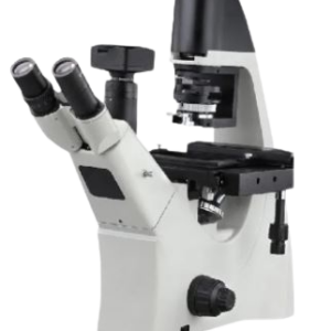
An inverted fluorescent microscope uses fluorescence to study specimens, such as living cells at the bottom of a petri dish or tissue culture. It is called “inverted” due to the arrangement of the microscope’s components, which are placed in inverted positions.
Sailent Features:
- Precise design
- Easy to install
- Long service life
- Ocular (eyepiece) lens.
- Objective turret or Revolver (to hold multiple objective lenses)
- Focus wheel to move the stage.
- Light source, a light or mirror.
Key Features:
- Anti Fungus Optics
- Optics With Multi Layers
- Easily Interchargable Objectives
- Choice Of Halogen And Led Illumination
- Main Operating Controls Within Easy Reach
- Optimization Of Illumination With Aspheric Lenses
- Available In Binocular And Trinocular Version
Scope Of Application
We know that the major uses of microscope is to view objects which are so tiny that they are invisible to naked eye. There are various applications of this devices depending on the fields it is used.
Uses of Microscope
- Botanical Field.
- Biological Field.
- Crime Investigation.
- Educational Field.
- Medical Field.
TECHNICAL DATA:
| PARAMETER | VALUES |
| Eye piece | Aberration free focusable 10x paired eyepiece lenses having field of view higher than 23/25mm (higher preferrable) |
| Microscope Frame | • Inverted microscope with full metal framesupporting with long lasting LED Fluorescence, Bright field,Phase contrast, DIC application facilities • System compatible to Micromanipulation facility, scanning stage upgradation. • Encoded 6-revoloving nosepiecewith provision for DIC facility • Facility for light manager system for transmitted light application. Microscope functions with camera via On screen display usage with automatic features for cameracontrol, Image enhancement function sand Readout toencoded microscope functions-Providedinterface |
| Transmitted Light | Transmitted white light LED with life span 60000 hrs. or more along with filter slider for enhancement filters ND 0.06 & balance filter.Microscope ECO mode facility to save energy of lamp. |
| Observation Tube | Phototube with 23/25mm field of view with 100% light distributionon camera/ ob servati on |
| Image analysis software | Image acquisition and processing software for extensive acquisitionand analysis facilities, multi-channel fluorescence, interactive measurement, vide file, multi-focusing etc. |
| Revolving Nosepiece | 6-revoloving encoded nosepieces with provision for (Differential Interference Contrast) DIC facility |
| Contrasting Methods | · Bright Field · Phase contrast · LED fluorescence |
| Condenser | Optically suitable matching long distancecondenser 0.4 mm with 53 mm working distance with 2.5x diffusion disc and as per supplied objective for bright field, phase contrast. |
| Stage | Microscope a hard coat anodized specimen stage to accommodate various specimen holders. It an object guide with long coaxial X-Y drive knobs and holders for various specimen containers like petridishes, tissue culture flasks, multiwellcell culture plates, glass slides etc. System up-gradation to scanning stage facility |
| Objectives | It 10X, 20X, 40X FL and 100X (for oil immersion) withfor fluorescence& phase contrast application with high NA. |
| Fluorescence illumination | Solid-State Light source for UV, Blue, Green excitation. 3-channel fluorescence light source with integrated control unit in stand for continuous brightness adjustment, quickly switchable and adjustable by stand alone, Onscreen Display (OSD) in camera software App. Equipped with 3 solid state LED lamps (25000Hr approx.). – Green (565nm) for excitation, – Blue (470nm) for excitation & – UV (385nm) for excitation. |
| Camera | High resolution monochrome CMOS/CCD HD camerawith minimum 2.Omegapixel with passive cooling, pixel size minimum 5.86pm x 5.86pm large pixel size with high sensitivity & automatically adjustable exposure& gain. Standalone camera capable of capturing good quality fluorescence imagewith PC & frame rate 30 fps, and high exposure range 0.3ms up to 2s Camera Image should have enhancement functions active denoising, active sharpening. |
| Computer | · A Branded desktop(HP) with minimum Intel Core i3 · 1TB HDD · 8GB RAM · 512 SSD · USB port · HDMI port · Licensed genuine Windows 10/11 (64 bit) OS · 20”LED monitor& UPS 15 min back up |
Accessories:
- Anti-fungal treated/Fungal treatment
- Lens cleaning kit
- lens cleaning paper
- immersion oil
- fiber box for microscope.





Reviews
There are no reviews yet.