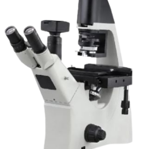
Inverted Fluorescent Microscope
MAKE DINESH SCIENTIFIC MODEL DS-IFM-10
PRODUCT DESCRIPTION
We offer a wide array of Inverted Research Microscope, which is easy to install and requires minimal maintenance.Designed under the supervision of skilled professionals, the offered products are appreciated by customers not only for their excellent performance and easy to use controls, but also for a good operational life. Further, we offer these products in various models.
SAILENT FEATURES:
- Precise design
- Easy to install
- Long service life
- Ocular (eyepiece) lens.
- Objective turret or Revolver (to hold multiple objective lenses)
- Focus wheel to move the stage.
- Light source, a light or mirror.
Scope Of Application
We know that the major uses of microscope is to view objects which are so tiny that they are invisible to naked eye. There are various applications of this devices depending on the fields it is used.
Uses of Microscope
- Botanical Field.
- Biological Field.
- Crime Investigation.
- Educational Field.
- Medical Field.
Illumination:
- Light source: LED (approx 5W or more) with intensity control, 50000 hrs life With Filter holder, built in filter, built in fly eye lens
- Condensor: Ultra long working distance condensor, N.A. 0.3 and above, W.D. 72mm, detachable
Photographic attachment:
- Colour Camera: 5 MP or more, CMOS sensor, 30 fps or more speed, wifi camera
- With image capturing software
- Microscope with camera & softwere
- Data transfer-HDMI, WLAN
- With voltage stabilizer having time delay function
- With vinyl dust cover
- 40 X objective
TECHNICAL DATA:
| PARAMETERS | VALUES |
| Type of Microscope | Infinity‐corrected optical system with Royal Microscopical Society (RMS) threaded objectives with a 45 mm par focal distance supporting fluorescence, bright field, color bright field, and phase contrast imaging modes |
| Compact integrated unit | Microscope, digital cameras, computer, high power fluorescence lighting system for Neurobiology, Immuno‐oncology Immunohistochemistry (IHC) applications etc,. |
| Illumination | Five‐position chamber for 4 fluorescence illuminators plus bright field imaging |
| Type illuminator of Light | Light illuminators with integrated hard‐coated filter set and LED light source with >50,000‐hour life |
| Broad selection of standard and specialty LED illuminators | Yes |
| Imaging methods | Single color, multicolor, time lapse, Z‐stacking, movie capture |
| Condenser | 60 mm LWD condenser; 4‐position turret with a clear aperture and 3 phase annuli |
| Stage | Mechanical X/Y stage |
| Travel range | 120 mm x 80 mm with sub micron resolution |
| Insert the holders | Drop‐in inserts to receive vessel holders and lockdown holders to fix sample in place |
| Include a universal holder and a multiwell plate holder | Yes |
| Focus mechanism | Automated focus mechanism with sub micron (0.150 um) resolution (single step accuracy) |
| Focus wheel | Mechanical focus wheel with single knob for coarse and fine focus |
| Position objective turret | 5 position objective turret with front mounted control |
| Plain Fluorite | 10X LWD PH‐ 0.25NA/9.2WD
20X LWD PH‐ 0.40NA/3.1WD, EACH 60X LWD‐ 0.75NA/2.2WD, EACH |
| Fluorite | 40X LWDPH 0.65NA/1.79WD, EACH |
| 05 independent high output LED illuminators | · DAPI, Ex: 357±2/44 nm; Em: 447±2/60 nm
· GFP, Ex: 470±2/22 nm Em: 510±2/42 nm · RFP, Ex: 531±2/40 nm; Em: 593±2/40 nm · Cy5, Ex: 628±2/40 nm; Em: 693±2/40 nm · Texas Red, Ex: 585±2/29; Em: 624±2/40 nm The LED illuminators will have independent intensity control |
| Installation of Illumination of light | Fluorescence LED illuminators single, interchangeable cubes that can be easily removed, installed and automatically recognized by the instrument software and adjust the configuration accordingly |
| Sensitivity | High ‐ sensitivity 3.2 MP (2,048 x 1,536) |
| Sensor | Monochrome CMOS sensor |
| Resolution pixel | 3.45 μm |
| Image Contrast Phase | 1click RGB channel overlay and also able to sequentially acquire a phase contrast image |
| Image fluorescence | Fluorescence image with a single mouse |
| User review for Microscope | Measure, and annotate captured images and count cells in fluorescence mode post acquisition |
| Perform Stem cell colony dissection with the fluorescence channels | Yes |
| Compatible with the onstage Incubator | Precise control of temperature, humidity, and gases for normoxic or hypoxic conditions allows a wide range of biological studies under physiological conditions |
| Microscope Software | Wizard based software and include downloadable software updates from time to time at |
| Connectivity | Windows/SMB network via an Ethernet cable connection and USB 3.0 WiFi dongle |
| Output file formats | 16 bit monochrome TIFF or PNG (12 bit dynamic range); 8 bit color TIFF, PNG JPG and BMP. |
| Output ports | Power, 4 USB 2.0 ports, 1 USB 3.0 port, 1 Display Port, 1 RJ45 network jack |
| Display | LCD Display ‐ 18.5″ articulated LCD color monitor with 1920×1080 pixel resolution |






Reviews
There are no reviews yet.