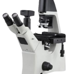
INVERTED FLOURESCENCE MICROSCOPE
MAKE DINESH SCIENTIFIC
DESCRIPTION
An inverted microscope is a specialized optical microscope where the light source and condenser are positioned above the stage, and the objectives are located below the stage. This design is the opposite of a standard microscope, hence the name “inverted.” It is primarily used to observe specimens in containers like petri dishes or culture flasks, which makes it ideal for studying live cells and organisms in their natural environment. Here are the key features:
Key Features:
Optics:
- High-quality objectives located below the stage, allowing the examination of thick samples or samples in dishes.
Stage:
- Fixed stage designed to hold large or irregularly shaped containers, such as culture dishes or flasks.
Illumination:
- Typically uses transmitted light from above, combined with fluorescence or phase contrast methods for enhanced imaging.
Applications:
- Ideal for live cell imaging, cell culture observation, and in vitro studies.
Magnification Range:
- Usually between 4x and 100x objectives.
TECHNICAL DETAILS:
| MODEL | DS-IFM-100 |
| Optical System | Infinity Optical System |
| Illumination | White LED diascope illumination with high brightness and an integrated “Fly Eye Lens” |
| Eyepiece | Pair of 10X (22mm FOV) |
| Body | Integrated diascopic illumination pillar, integrated LED illumination, and an objective elevation system for focusing are all features of the inverted microscope body. Diascopic/Epifluorescence switching controls and an ON/OFF switch are located in the front panel. |
| Stage | XY stage of 170 x 240 mm with a moving range of 125(X) x 75(Y) mm |
| Holder | Universal holder for Terasaki plate holder |
| Tube | Binocular Tube inclined at 45°, pupillary distance 50-75mm |
| Camera Port | Attachable camera port, Eyepiece/Port- 100/0: 0/100 |
| Nose Piece | Quintuple Nosepiece |
| Fluorescence Filter | UV and FITC fluorescence filters, respectively. |
| Upgradability | Upgradable to add more filters |
| Condenser | Long working distance condenser 75mm , N.A. 0.3 |
| Observation | Brightfield, Phase Contrast & Epifluorescence |
| Objectives | Plan Achromat 4X, Plan Achromat LWD DL 10X, Plan Achromat LWD DL 20X, Plan Achromat LWD DL 40X, Plan Achromat LWD DL 100X |
| Epifluorescence Attachment | LED Fluorescence Filter turret with 3 positions |
| SOFTWARE | |
| Software Compatibility | Suitable for taking pictures, compatible with the microscope, and equipped with extra features. |
| Basic Measurement Tools | Includes tools for basic measurements during analysis. |
| Real-Time Fluorescence Synthesis | Enables real-time fluorescence synthesis and editing to improve utility. |
| Single-Shot Imaging | Allows for precise single-shot imaging using a delayed camera. |
| Time-Lapse Photography | Allows for time-lapse photography to capture changes over time. |
| Z-Profile Generation | Capable of generating z-profiles for analyzing the image in 3D. |
| Multichannel Imaging | Integrates multichannel imaging capabilities for capturing multiple image channels simultaneously. |
| HDR Image Synthesis | Supports HDR image synthesis for enhanced dynamic range and image quality. |
| Image Stitching | Makes it possible to stitch several photos together to create a single, high-resolution image. |
| Annotation Features | Includes annotation tools to mark and label images for further analysis and reference. |
| CAMERA | |
| Image sensor | Colour CMOS |
| Resolution | 6MP |
| Pixel Size | 2.4 pm x 2.4pm |
| Bit depth | 8bit |
| Exposure settings | Auto/Manual |
| Data Interface | USB 3.0 |
| Mount | C mount |
| Exposure time | 15us – 10s |
| Picture format | JPG / PNG / TIFF / compatible with MS-Office |





Reviews
There are no reviews yet.