FLUORESCENCE MICROSCOPE
MAKE DINESH SCIENTIFIC
DESCRIPTION
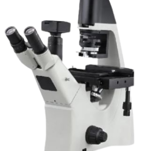
A specialized kind of optical microscope called an inverted fluorescence microscope is used to view and photograph specimens that glow when exposed to particular light wavelengths. An inverted fluorescence microscope differs from conventional microscopes in that the objective lens and detector are located below the specimen stage, while the light source and condenser are located above it. This shape makes it more practical and convenient to examine thicker objects, including living animals or cell cultures.
LIGHTING FRAMEWORK:
Usually outfitted with a strong light source, like a xenon or mercury lamp, that may emit particular light wavelengths that are ideal for stimulating the specimen’s fluorophores.
- Include a filter wheel or cube to choose the right excitation wavelength for the fluorophores being employed.
SYSTEM OF CONDENSERS:
- Intended to concentrate and direct the excitation light onto the specimen, it is positioned above the stage.
- Contains lenses and filters frequently to improve the excitation light’s quality and specificity.
OBJECT LENSES:
- These lenses, which are situated beneath the specimen stage, enlarge the fluorescence that the specimen emits.
- To obtain finely detailed, high-resolution images, high numerical aperture (NA) objectives are frequently utilized.
STAGE OF SPECIMEN:
- Thick specimens, such as cell cultures on petri dishes or multi-well plates, can be examined with the setup inverted.
- Frequently outfitted with a system for regulating humidity and temperature to keep live-cell imaging conditions ideal.
FILTERS FOR FLUORESCENCE:
- Positioned to block the excitation light and selectively transmit the specimen’s fluorescence while it is in the path of light.
- Multiple fluorophores can be seen at once by using different filter sets.
TECHNICAL DETAILS:
| MODEL | DS-FM-100 |
| Microscope Type | Upright type fluorescence microscope with photographic attachment |
| Unit Composition | The microscope is a small, integrated instrument that includes a computer system, a high-performance digital camera, a cell imaging system, and a high-performance fluorescent illumination system. |
| MICROSCOPE TYPE | |
| Microscope Type | Research-grade trinocular featuring an easy-to-use lamp replacement feature and an integrated light and intensity regulator. |
| Illumination | LED illumination for transmitted light with a lamp life of 50,000 hours |
| Objective Nosepiece | Revolving nosepiece with 7 positions |
| Observation Method | Bright field and fluorescence |
| Observation Tube | Trinocular tube with 0/100:100/0 beam splitter for optimal signal acquisition |
| Objective Types | · Plan Achromatic
· Plan Achromatic Infinity · Semi Apochromatic |
| Objective Magnifications and Numerical Aperture | 5X/0.12, 10X/0.25 (Phase Contrast), 40X/0.65 (Phase Contrast), 100X/1.25 (oil immersion) |
| Transmission Efficiency | For efficient fluorescence microscopy, each objective has a transmission efficiency of 80%. |
| Condenser | For bright field and fluorescence applications, a universal condenser with color coding allows for quick and simple aperture diaphragm adjustment. |
| Eyepiece Type | Compensating eyepiece |
| Eyepiece Magnification | Set of two binocular eyepieces with 10X magnification and a field of view (FoV) of 22 mm |
| Photographic Unit | High-resolution cooled color CMOS camera with 6 MP resolution |
| Stage of the Microscope | Rectangular shape, rotatable, measuring 75 mm x 50 mm |
| Stage Movement | Coarse and fine movement present |
| Focusing Arrangement | Coaxial focusing and micrometer arrangement present |
| Interface | USB 3.0 interface |
| Image Capture Capability | Capable of capturing images for both bright-field and fluorescence microscopy. |
| Image Acquisition Rate | 15 frames per second |
| FLUORESCENCE ILLUMINATION | |
| Fluorescence Illumination | Five-position fluorescence filter turret; intense 100-watt Hg fluorescent illumination. |
| Spectrum Coverage | Broad spectrum covering the UV region (DAPI filter) to the red region (Cy5 filter) |
| FLUORESCENCE FILTER DETAILS | |
| DAPI/Hoechst | · Excitation: BP 350/50
· Dichroic: LP 400 · Emission: BP 460/50 |
| FITC/GFP | Excitation: BP 480/40, Dichroic: 505, Emission: 527/30 |
| Rhoda mine/TRITC/Cy3 | Excitation: BP 515-560, Dichroic: 580, Emission: LP 590 |
| Software Specification | Licensed imaging program for spatial measurements (length, width, area, perimeter, etc.), fluorescence multichannel overlay, intensity measurements, counting, and line profiles. |
| DATA PROCESSING UNIT | |
| Data Processing Unit | Branded PC with compatible processor |
| RAM | 16 GB |
| Monitor Size | 24 inches for efficient image analysis |
| Graphics Card | NVIDIA graphics card with 2 GB RAM |
| Storage | 1 TB SATA HDD |
| Operating System | Installed with original (licensed) compatible operating system |
| Additional Features | DVD RW function |
| Power | Functional with standard plug points (220-240 VAC, 50/60 Hz, 1 phase) |





Reviews
There are no reviews yet.