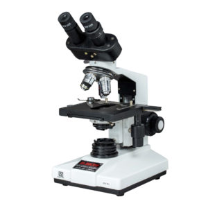
BRIGHT FIELD OPTICAL MICROSCOPE
MAKE DINESH SCIENTIFIC
MODEL DS-MS-100
DESCRIPTION
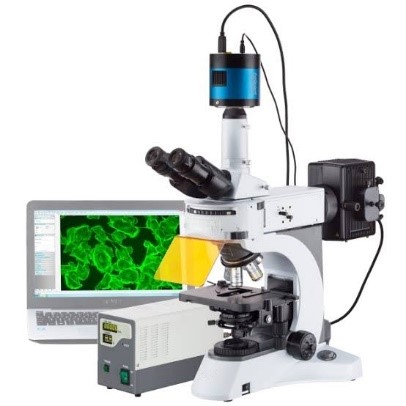
An inverted microscope is a specialized optical microscope where the light source and condenser are positioned above the stage, and the objectives are located below the stage. This design is the opposite of a standard microscope, hence the name “inverted.” It is primarily used to observe specimens in containers like petri dishes or culture flasks, which makes it ideal for studying live cells and organisms in their natural environment. Here are the key features:
Key Features:
Optics:
High-quality objectives located below the stage, allowing the examination of thick samples or samples in dishes.
Stage:
Fixed stage designed to hold large or irregularly shaped containers, such as culture dishes or flasks.
Illumination:
Typically uses transmitted light from above, combined with fluorescence or phase contrast methods for enhanced imaging.
Applications:
Ideal for live cell imaging, cell culture observation, and in vitro studies.
Magnification Range:
Usually between 4x and 100x objectives.
Focus Mechanism:
Coarse and fine focus adjustments for precise imaging.
TECHNICAL DETAILS:
MODEL DS-MS-100
Observation Method Inverted microscope with Bright field, Phase contrast, Fluorescence (Ultraviolet Excitation and Blue, Green Excitation).
Phase Ring Phase ring for 10X and 40X.
Fluorescence Fluorescence attachment for 6 cube along with narrow band pass filters for DAPI, FITC/GFP, and TRITC filters.
Fluorescence Light Source 100-watt mercury burner or LED light source for fluorescence imaging.
Microscope Frame Dual deck frame with high static rigidity, waterproof construction. Compact design with frontal controls for light path selector (0:100/50:50/100:0), transmitted light intensity control, and light ON/OFF switch. Comes with sextuple revolving nosepiece with DIC slider.
Optical Lenses All optical lenses UV-IR corrected.
Observation Tube Binocular observation tube with diopter adjustment on both eyes.
Eyepiece 10X eyepiece with F.N. 22 and diopter.
XY Stage Rectangular mechanical XY stage with right-hand long type control handle, universal sample holder for large plates, glass slides, petri plates, etc.
Condenser Universal long working distance condenser with 5 positions for optical elements, working distance of 27-28 mm.
Transmitted Light Illuminator LED-based transmitted light illumination pillar with tilt mechanism, condenser holder, field iris diaphragm, and 4 filter holders.
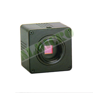
Camera • High-resolution CMOS camera for fluorescence samples and live cell imaging.
• High-resolution, color CMOS Camera chip.
• 8.9-megapixel with 4K resolution
• Pixel size: 2.4 µm² ,FOV 25MM.
• Live display: 64 FPS.
• Seamless integration of camera and software
Image Acquisition and Analysis Software Software for image capturing, automated FL image merging, overlay multiple images, movie playback, tile imaging, time-lapse datasets, manual and live deblurring, image processing, image analysis, basic measurements (phases/regions), automated multi-wavelength, region and line measurement, interactive measurement, counting.
Power Supply Compatible with 220 V, 50 Hz power supply.
UPS A compatible UPS provided for the instrument.
Objectives • Universal Plan Fluor/Plan Semi Apochromat Phase Objective 10X/0.3, WD 10mm.
• Long working Plan semi-Apochromatic objective 40X phase, NA 0.60, with correction collar.
• Plan Semi Apochromat 100X/1.30 objective, Oil.
• Plan Fluorite 100X/0.95, WD 0.2 Objective Dry.
Computer and Software • Genuine Windows OS
• Intel Core i5 processor
• 4GB RAM
• 1TB HDD
• DVD R/W
• 24’’ LCD/TFT monitor
• Compatible 1 KVA offline UPS for microscope & camera control.
Saving your money for next purchases
Delivery – when you want and anywhere





Reviews
There are no reviews yet.