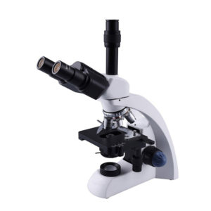
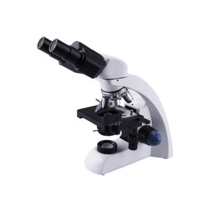
INVERTED MICROSCOPE
MAKE DINESH SCIENTIFIC
DESCRIPTION:
Because they allow researchers and scientists to examine tissues, cells, and other biological material at a microscopic level, microscopes are essential tools in pathology and medical science. Research and pathology microscopes are made to specifically address the needs of the medical, biological, and allied sciences sectors. The following are some of the of the essential elements of using microscopes in research and pathology settings:
LIGHT MICROSCOPES:
- The most popular kind of microscope used in pathology and research are brightfield microscopes. They are useful for seeing stained specimens and offer a bright background.
PHASE-CONTRAST MICROSCOPES:
- When viewing translucent or unstained materials, such as living cells, phase-contrast microscopy is especially helpful. It improves these specimens’ contrast without the requirement for staining.
FLUORESCENCE MICROSCOPES:
- Research uses fluorescence microscopy extensively. It entails labeling certain molecules or structures within cells with fluorescent dyes or proteins to enable fine-grained imaging of individual cell components.
PATHOLOGY DIGITAL:
- Digital imaging technology is used in the collection, organization, and analysis of pathology data in digital pathology. Pathologists can see and evaluate digital slides using whole-slide imaging, which facilitates remote diagnosis and teamwork.
MECHANIZED MICROSCOPY:
- Microscopy technologies are becoming more and more automated, enabling higher throughput analysis and more productivity in pathology and research labs.
RESEARCH USES:
- In several scientific fields, such as cell biology, microbiology, neuroscience, and developmental biology, microscopes are essential instruments. The structure and operation of cells, tissues, and organisms are studied by researchers using microscopes.
TECHNICAL DETAILS:
MODEL |
DS-IM-10 |
| Microscope stand | Furnished with an LED transmitted-light and a quadruple rotating nosepiece |
| Mechanical stage | Has a movable right-hand low drive control and plate and glass slide holders. |
| Koehler Illumination | Koehler lighting is appropriate for phase contrast and bright field microscopy |
| Optical system | To optimize optical performance and signal-to-noise ratio from UV to near infrared, infinity correction is applied. |
| Phase contrast slider | Phase contrast applications are pre-centered, including positions for 4X, 10X, 20X, and 40X objectives. |
| LED illuminator | Offers a lifespan of 20,000 hours |
| Condenser | A long-working-distance condenser measuring 72 mm with a 0.3 numerical aperture |
| Data transfer | Supports HDMI, USB 2.0, WLAN via internet, and SD card |
| Digital camera | 5MP color CMOS sensor with PC and HDMI interface and a high resolution of 2592 x 1944 pixels |
| Eyepieces | 10X magnification with adjustable diopter |
| Objectives | Plan-Achromat Phase Contrast Long Working Distance Objectives: 4X, 10X, 20X, and 40X |






Reviews
There are no reviews yet.