POLARIZING MICROSCOPES
MAKE DINESH SCIENTIFIC
DESCRIPTION
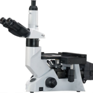
A standard polarizing microscope is equipped with essential features for routine polarized light microscopy. It typically includes a transmitted light base with a rotatable stage for easy specimen orientation. The microscope has a built-in Bertrand lens and compensator slot for analyzing birefringent materials. The optical system ensures clear and high-contrast images, making it suitable for basic geological and materials science applications. Standard polarizing microscopes often come with a set of objectives, eyepieces, and polarizing filters to facilitate straightforward sample analysis.
LIGHT MICROSCOPES:
- The most popular kind of microscope used in pathology and research are brightfield microscopes. They are useful for seeing stained specimens and offer a bright background.
RESEARCH-GRADE POLARIZING MICROSCOPE:
- A research-grade polarizing microscope is designed for advanced applications in materials science, geology, and other research fields. It features high-quality optics with advanced lens coatings to enhance image resolution and clarity. The microscope often includes a modular design, allowing for customization based on specific research needs. Research-grade polarizing microscopes come with a range of objectives, compensators, and imaging accessories to provide comprehensive analytical capabilities. These microscopes may also have motorized components for automated imaging and analysis.
PATHOLOGY DIGITAL:
- Digital imaging technology is used in the collection, organization, and analysis of pathology data in digital pathology. Pathologists can see and evaluate digital slides using whole-slide imaging, which facilitates remote diagnosis and teamwork.
MECHANIZED MICROSCOPY:
- Microscopy technologies are becoming more and more automated, enabling higher throughput analysis and more productivity in pathology and research labs.
RESEARCH USES:
- In several scientific fields, such as cell biology, microbiology, neuroscience, and developmental biology, microscopes are essential instruments. The structure and operation of cells, tissues, and organisms are studied by researchers using microscopes.
TECHNICAL DETAILS:
| MODEL | DS-APM-X |
| Condenser | POL 0.85 with lambda % compensator |
| LED Illumination with attachments for TL | Includes all attachments for transmitted light (TL) applications. |
| A/B module | Advanced colposcopy module with focusable and central Bertrand lens |
| Polarizing stage with object guide | Object guide for Pol-Stages with xy-control, suitable for different slide formats |
| · 4 Position enterable nosepiece.
· Koehler Illumination. · Circular rotating stage (Diameter > 175 mm) with graduations and adjustable brake. · LED illumination. · Built-in handle and cord wrap. · Objective centering tools. · 2 Object Clamps. · Dust Cover. · USB 2.0 interface for connection to a PC including USB 2.0 cable. |
|
| Observation Tube | ü 30-degree Polarization Tube with eyepiece locking mechanism.
ü Orientation key in one eye tube for the Crosshair reticule eyepiece |
| Eyepiece with eye guard | · 10x focusable eyepiece with Crosshair reticule
· Key for orientation |
| Objectives | Pol4x/0.10, Pol10x /0.25, Pol20x /0.40, Pol40x/0.65, Free working distance suitable for use with and without cover glass for all magnifications, Suitable for TL applications and upgradable for reflected light application. |
| Microscope’s possibility to upgrade with camera | Ability to upgrade with Digital Imaging camera system |
| OPTIONAL | |
| Future upgrade All apochromatic lenses | 0.5x/0.63x/0.75x/2.0x
|
| IMAGING SOFTWARE | |
| Imaging software for provided with the microscope. It capable of -Image Processing & Measurement: | · Modify each image’s gamma, contrast, and brightness
· Combine, crop, and do image arithmetic . · Area intensities are measured using picture stacking · Parallax adjustment. |
| Camera, and software | For improved synchronization, choose a single manufacturer for the microscope, camera, and software. |
| One Camera | High-speed digital CMOS camera with 12 MP. 4K HD video at 30 or 60 frames per second. recording at 30 frames per second at 1080p. ability to attach a camera by Ethernet, HDMI, or WiFi. Without a PC, images can be transferred directly to a USB stick. Without a PC, standalone operation is feasible. CMOS sensor (1/2.3), pixel size 1.55 x 1.55 pm, exposure time 100 ps – 120 ms. 24 bit color depth. Offers a 0.55x C mount. |
ACCESSORIES AND MANUALS:-
- Operating manuals.
- Sufficient sheets of adhesive labels for 6 function keys for all microscopes.
- Microscope covers (with 3 spare pieces) provided.






Reviews
There are no reviews yet.