")
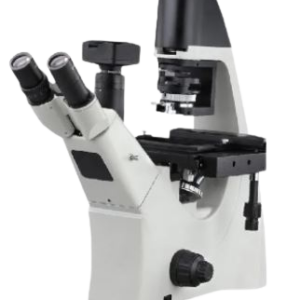


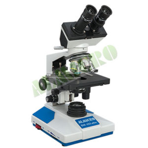
Inverted Research Microscope
Make Dinesh Scientific
Model DS-IRM-100
PARAMETER VALUE
SALIENT FEATURES:
Compact tabletop design (≤ 14” footprint)
Multi-mode observation: Brightfield, Darkfield, Phase Contrast, and
Fluorescence
Advanced transmitted light source: White LED or 12V 100W Halogen
with intensity control
Dual port light sharing: 100:0 and 0:100 configurations (camera:eye)
Universal phase contrast condenser compatible with all imaging
modes
High precision manual X-Y-Z axis stage with universal sample holder
Five-position objective nosepiece for flexibility in magnification
High-performance objectives with wide working distance and high
NA
DESCRIPTION:
The is a compact, portable inverted fluorescent microscope designed for high-performance imaging in brightfield (BF), darkfield
(DF), phase contrast (PC), and fluorescence (FL) modes. With a minimal footprint not exceeding 14 inches, it is ideal for spaceconstrained
laboratories and routine imaging needs. Engineered for flexibility and precision, it integrates a universal phase contrast
condenser, ergonomic manual XYZ stage, and advanced imaging capabilities suitable for research and diagnostics. The microscope
supports dual light paths (100:0, 0:100) between the camera and eyepiece ports and is optimized with high-grade objectives,
including Semi-Apochromat/Neo Fluor/Plan Fluor lenses.
Feature Category Specifications / Description (Rephrased)
Microscope Type Compact, portable tabletop inverted fluorescent microscope
with a footprint not exceeding 14 inches
Observation Methods Equipped for Bright Field (BF), Dark Field (DF), Phase
Contrast (PC), and Fluorescence (FL) imaging
Image Analysis Integrated advanced image analysis capabilities, with
preference for systems supporting dark field imaging
Illumination Advanced transmitted light source, either high-intensity
white LED or 12V 100W halogen, with adjustable brightness
Light Sharing Dual-mode light path switchable between camera and
eyepiece ports (100:0 and 0:100 distribution)
Condenser Universal phase contrast condenser compatible with all
observation techniques
Nose-piece Quintuple (5-position) objective nosepiece for flexible lens
configuration
Stage Manual X, Y, and Z axis mechanical stage featuring a
universal holder for various sample types
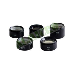
Objectives Includes 4X, 10X, 20X, 40X, and 100X (oil immersion)
objectives optimized for BF, DF, PC, and FL imaging modes
TECHNICAL SPECIFICATION:
Objective Details
4X/5X Semi-Apochromat / Neo Fluor / Plan Fluor; NA 0.13,
WD 17 mm; for BF, DF, and FL modes
10X Semi-Apochromat / Neo Fluor / Plan Fluor; NA 0.30,
WD 10 mm; suitable for BF, DF, PC, and FL
20X Semi-Apochromat / Neo Fluor / Plan Fluor; NA 0.45,
WD 6.6–7.8 mm; for BF, DF, PC, and FL
40X Semi-Apochromat / Neo Fluor / Plan Fluor; NA 0.60,
WD 3.0–2.5 mm; for BF, DF, PC, and FL
100X (Oil) Semi-Apochromat / Neo Fluor / Plan Fluor; NA
1.30, WD 0.16 mm; compatible with BF, DF, and FL
Eyepiece Pair of 10X widefield eyepieces with 22 mm field of view
Accessories Supplied with suitable immersion oil in small-volume bottles
(50 ml each)
Fluorescence module
Fluorescence Turret Type Available in manual, coded, or intelligent configurations for
flexible fluorescence filter management
Filter Positions Equipped with a 4-position turret, accommodating three
active filters and one additional/reserve slot
Functionality Allows users to define and store specific LED illumination
settings for each fluorescence channel
Wavelength Memory Feature Automatically retrieves and applies the pre-set illumination
intensity when a previously used wavelength is selected
Camera
Camera Type High-performance scientific-grade color digital camera for
microscopy applications
Connectivity Equipped with USB 3.0 interface ensuring fast data transfer
and seamless PC/laptop integration
Imaging Compatibility Supports imaging modalities including Fluorescence, Phase
Contrast, Dark Field (DF), DIC, and Bright Field
Sensor Type Utilizes a high-quality CMOS sensor
Sensor Size 1/1.8-inch sensor format
Resolution Preferred resolution range of 15 to 20 megapixels for detailed
image capture
Pixel Size Pixel dimensions of 3.45 μm × 3.45 μm for enhanced image
clarity and sensitivity
Bit Depth Offers 12-bit image depth for accurate color and grayscale
rendering
Live Display Speed Capable of delivering live video at 25–30 frames per second
(FPS) at 1K resolution
Accessories Supplied complete with all required accessories for image
acquisition and transfer to a laptop or PC
Fluorescence Light Source
Illumination Type LED-based fluorescence illumination system
LED Lifespan operational life of 10,000 hours
Filter Sets Included Pre-installed with DAPI, FITC, and TRITC filter sets
Filter Upgrade Capability Compatible with and upgradeable to advanced or additional
fluorescence filter sets as required
Image capturing and storing device
Computer Type Lightweight and portable laptop system suitable for
laboratory and imaging applications
Processor Equipped with Intel i7 processor for high-speed performance
Storage Integrated 1TB hard disk drive for ample data storage
Memory (RAM) 12 GB RAM to ensure smooth multitasking and software
operation
Display High-resolution TFT color display, available in either 15” or 21”
screen size
Operating System Pre-installed with genuine licensed operating system
Office Suite Comes with licensed multi-user Microsoft Office software
Antivirus Software Supplied with multi-user licensed antivirus protection
Keyboard Includes multimedia keyboard for enhanced usability
Graphics Card Features a dedicated 1GB graphics card for improved image
and video rendering
System Unit System cabinet/unit not included as part of the supply
Microscope Accessories Includes a 32 GB USB pen drive and a 1TB external hard disk
for data backup and portability
Software
Software Type Advanced imaging software for integrated control of
microscope, illumination, and camera components
Control Functions Enables complete control of microscope hardware, LED
illumination, and camera functionalities
Key Features
Multi-channel image acquisition
Large image stitching with reference mirror image
Time-lapse imaging
Multi-channel fluorescence imaging
Volume rendering and visualization
2D and 3D image viewing
Morphological analysis
Automatic object counting
Automated measurements
Z-stacking for depth imaging


Reviews
There are no reviews yet.