")
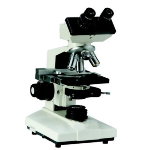


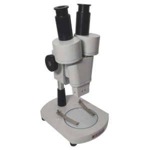
MICROSCOPES – PATHOLOGICAL AND RESEARCH
MAKE DINESH SCIENTIFIC
MODEL DS-BM-100
DESCRIPTION
Because they allow researchers and scientists to examine and examine tissues, cells, and other biological
material at a microscopic level, microscopes are essential tools in pathology and medical science. Research
and pathology microscopes are made to specifically address the needs of the medical, biological, and allied
sciences sectors. The following are some essential elements of using microscopes in research and pathology
settings:
LIGHT MICROSCOPES:
✓ The most popular kind of microscope used in pathology and research are
brightfield microscopes. They are useful for seeing stained specimens and
offer a bright background.
PHASE-CONTRAST MICROSCOPES:
✓ When viewing translucent or unstained materials, such living cells, phasecontrast
microscopy is especially helpful. It improves these specimens’
contrast without the requirement for staining.
FLUORESCENCE MICROSCOPES:
✓ Research uses fluorescence microscopy extensively. It entails labeling certain molecules or structures
within cells with fluorescent dyes or proteins to enable fine-grained imaging of individual cell
components.
PATHOLOGY DIGITAL:
✓ Digital imaging technology is used in the collection, organization, and analysis of pathology data in
digital pathology. Pathologists can see and evaluate digital slides using whole-slide imaging, which
facilitates remote diagnosis and teamwork.
CATALOGUE
TECHNICAL DETAILS:
Optical System Universal Infinity Corrected Optical System.
Nosepiece Inward-facing quadruple nosepiece.
Stage Wire movement mechanical fixed stage (W x D: 174mm
x 89mm), with a traveling range of 76mm x 30mm (X x
Y).
Illuminator Integrated, 20,000-hour-lasting transmission LED
lighting.
Focusing Stage height movement with a coarse movement stroke
of 15mm, torque adjustment for coarse focusing knob,
coarse and fine focus knobs on both sides, and an upper
limit stopper to prevent objective lens from hitting the
slide.
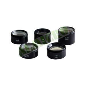
Objectives Completely field-flat Plan Achromat objectives:
• 4X (N.A. 0.10, WD 27.8mm)
• 10X (N.A. 0.25, WD 8.0mm)
• 40X (N.A. 0.65, WD 0.6mm)
• 100X (N.A. 1.25, WD 0.13mm)
Eyepiece Paired 10X eyepieces with diopter adjustment and field
number 20mm, fixed in place to prevent damage.
Observation Tube An eyepoint height system for user comfort, a field
number of 20 mm, an interpupillary distance of 48-75
mm, and a Siedentop-type binocular observation tube
angled at 30°.
Cover Yes
Condenser Abbe condenser (N.A. 1.25) with built-in aperture
diaphragm for brightfield and darkfield.


Reviews
There are no reviews yet.