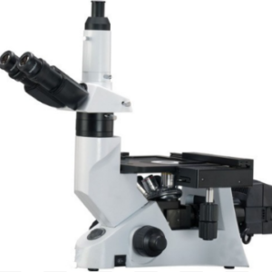
INVERTED MICROSCOPE
MAKE DINESH SCIENTIFIC
DESCRIPTION
Because they allow researchers and scientists to examine and examine tissues, cells, and other biological material at a microscopic level, microscopes are essential tools in pathology and medical science. Research and pathology microscopes are made to specifically address the needs of the medical, biological, and allied sciences sectors. The following are some essential elements of using microscopes in research and pathology settings:
LIGHT MICROSCOPES:
The most popular kind of microscope used in pathology and research are brightfield microscopes. They are useful for seeing stained specimens and offer a bright background.
PHASE-CONTRAST MICROSCOPES:
- When viewing translucent or unstained materials, such living cells, phase-contrast microscopy is especially helpful. It improves these specimens’ contrast without the requirement for staining.
FLUORESCENCE MICROSCOPES:
- Research uses fluorescence microscopy extensively. It entails labeling certain molecules or structures within cells with fluorescent dyes or proteins to enable fine-grained imaging of individual cell components.
PATHOLOGY DIGITAL:
- Digital imaging technology is used in the collection, organization, and analysis of pathology data in digital pathology. Pathologists can see and evaluate digital slides using whole-slide imaging, which facilitates remote diagnosis and teamwork.
MECHANIZED MICROSCOPY:
- Microscopy technologies are becoming more and more automated, enabling higher throughput analysis and more productivity in pathology and research labs.
RESEARCH USES:
- In several scientific fields, such as cell biology, microbiology, neuroscience, and developmental biology, microscopes are essential instruments. The structure and operation of cells, tissues, and organisms are studied by researchers using microscopes.
TECHNICAL DETAILS:
| MODEL | DS-IM-20 |
| Imaging Channels | 4 channels: 1 bright field and 3 fluorescent channels |
| Blue Channel | · Excitation : 355/40 nm
· Emission : 433/36 nm |
| Red Channel | · Excitation : 556/20 nm
· Emission : 615/61 nm |
| Green Channel | · Excitation : 480/17 nm
· Emission : 517/23 nm |
| Illumination Light Sources | |
| Blue Channel | UV LED |
| Green Channel | Blue LED |
| Red Channel | Green LED |
| Bright Field Channel | Green LED for image contrast optimization |
| Imaging Channels | 4 channels: 1 bright field and 3 fluorescent channels |
| Screen Resolution | 1280 x 768 pixels |
| Tilt Range | 80-180° angle |
| Touch Screen | 10 inch LCD monitor |
| Screen Features | Touch screen with anti-fingerprint and anti-glare coating. |
| Image File Types | JPEG, TIFF, RAW |
| Image Capture | · Monochrome camera
· 12-bit CMOS · 5 megapixels |
| Focusing Mechanism | · Coarse and fine
· Manual adjustment |
| Compatible Consumables | · Flasks (T25, T75, T225), multi-well plates (6, 12, 24, 48, 96, 384 wells), dishes (35mm, 60mm, 100mm), chamber slide, glass microscopy slide |
| Numerical Aperture | 0.40 |
| Field of View (FOV) | 0.70 mm² |
| Data Export | 2 USB ports |
| Image Merge | Bright field and 3 fluorescent images can be overlaid |
| Data Storage | 2000 images, 16 GB internal memory |
| Objective | 20X |
| Magnification | · Display: 170X
· Digital Zoom: 700X |
| On-Board Software | Yes, Android operating system |
| Stand-Alone Operation | PC is not required for operation |
| Motorized Stage | 6 mm travel in X, Y direction
Touch screen control of travel speed and direction. |








Reviews
There are no reviews yet.