MICROSCOPE PATHOLOGICAL AND RESEARCH
MAKE DINESH SCIENTIFIC
DESCRIPTION
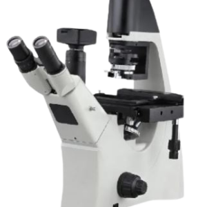
Because they allow researchers and scientists to examine and examine tissues, cells, and other biological material at a microscopic level, microscopes are essential tools in pathology and medical science. Research and pathology microscopes are made to specifically address the needs of the medical, biological, and allied sciences sectors. The following are some essential elements of using microscopes in research and pathology settings:
LIGHT MICROSCOPES:
- The most popular kind of microscope used in pathology and research are brightfield microscopes. They are useful for seeing stained specimens and offer a bright background.
PHASE-CONTRAST MICROSCOPES:
- When viewing translucent or unstained materials, such living cells, phase-contrast microscopy is especially helpful. It improves these specimens’ contrast without the requirement for staining.
FLUORESCENCE MICROSCOPES:
- Research uses fluorescence microscopy extensively. It entails labeling certain molecules or structures within cells with fluorescent dyes or proteins to enable fine-grained imaging of individual cell components.
PATHOLOGY DIGITAL:
- Digital imaging technology is used in the collection, organization, and analysis of pathology data in digital pathology. Pathologists can see and evaluate digital slides using whole-slide imaging, which facilitates remote diagnosis and teamwork.
MECHANIZED MICROSCOPY:
- Microscopy technologies are becoming more and more automated, enabling higher throughput analysis and more productivity in pathology and research labs.
RESEARCH USES:
- In several scientific fields, such as cell biology, microbiology, neuroscience, and developmental biology, microscopes are essential instruments. The structure and operation of cells, tissues, and organisms are studied by researchers using microscopes.
TECHNICAL DETAILS
| MODEL | DS-MS-10 |
| Microscope Type | pathological non-hinging binocular type with integrated light and adjustable light intensity. |
| LCD Screen | 32” |
| Binocular Eye pieces | yes |
| Size of Stage | 120X132mm |
| Eye Piece with magnification | Set of Two For Binocular 10x |
| Objective Type | Plan Achromatic Infinity |
| Eye piece Type | Compensating |
| Type of lamp for Illumination | led |
| Stage | Ractangular |
| Coarse and Fine Movement of stage | yes |
| Co-axial focussing | Yes |
| Objective Magnification | 10 x,20 x,40 x,4X |
| Numerical Aperture of Objective | 0.25,0.5,0.65,0.10 |
| ADDITIONAL | |
| Microscope Type | Upright |
| Illumination | Constant color LED illumination (5 Watt) with a life of 50,000 hrs., variable Kohler illumination |
| Magnification | Upgradable to PH, DIC, darkfield, fluorescence |
| Color Temperature | Maintained constant at all magnifications |
| Focusing | Coaxial course, fine focusing with torque and height adjustment focus knob, adjustable focus stop, 25mm Z travel range |
| Nosepiece | 6 positions |
| Miscellaneous | Upgradable to fluorescence with 5 position Fluorescence filter turret. Microscope, camera, and software included |
| Condenser | Color coded condenser suitable for BF, DF, Phase contrast |
| Objectives | HiPlan 4x/0.10, HiPLAN 10x/0.25, HiPLAN 20x/0.40, HiPLAN 40x/0.65, HiPLAN 100x/1.25 Oil |
| Eyepiece | Harmonic corrected, dioptre adjustable eyepiece 10x/20mm. |
| Binocular Tube | 30-degree angle |
| Camera | · Body-integrated color CMOS camera with 12 MP resolution, 60 frames per second, and support for 4K Ultra HD, USB3, HDMI, and USB Stick.
· Software for recording videos, capturing images, and operating on its own focus feedback, gallery, histogram · Save a picture to a network. Observation-based live measurement, image comparison, overlay, snap-to-edge support for circular measurements and annotations · High dynamic range Automated camera settings adjustment · Use Negative mode to accentuate details that are difficult to see. |
| Stage | · Ergonomic XY stage with coaxial xy drive, ultrahard ceramic surface with Vernier reading
· Right & left-hand operation, slide holder with one hand operation, LCD Display provided |








Reviews
There are no reviews yet.